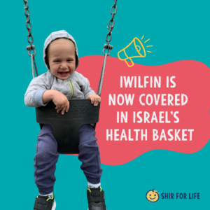
A new drug just entered Israel’s health basket. Here’s why it matters
A new drug just entered Israel's health basket. Here's why it matters The decision to add IWILFIN to the health ...
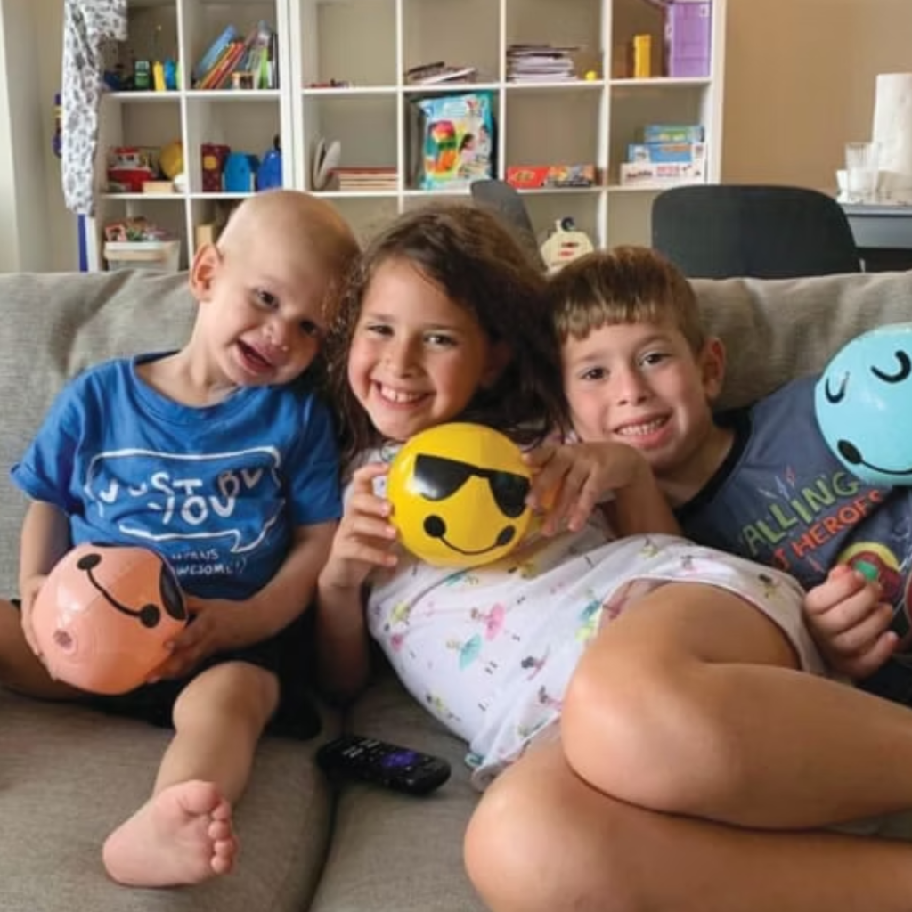
Why Collaboration, Not Competition, Will Cure Rare Cancers
The battle against neuroblastoma, the rare and deadly child-killing cancer By Einat Dado BaraliaThe Jerusalem PostDecember 5, 2025 When your child ...
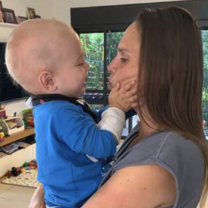
‘A Life-Changing Song’: Turning grief into hope for children with cancer
'A Life-Changing Song': Turning grief into hope for children with cancer By Einat Dado BaraliaYnetnewsDecember 17, 2024When Shir was diagnosed ...
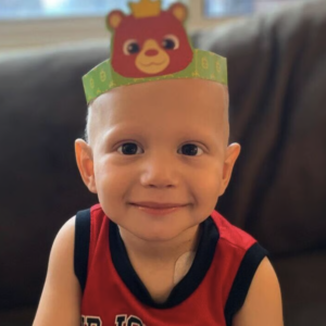
Remembering Shir: Lighting the Path Forward for Childhood Cancer Fighters
Every Child Deserves to Dream: International Neuroblastoma Awareness Day Is Here By Jerusalem Post Staff, August 23, 2023 Aug 23, ...